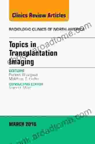
Topics in Transplantation Imaging: An Issue of Radiologic Clinics of North America

This issue of Radiologic Clinics of North America focuses on Topics in Transplantation Imaging. The issue editor is Dr. Richard C. Semelka, Professor of Radiology at the University of North Carolina School of Medicine in Chapel Hill, North Carolina.
5 out of 5
| Language | : | English |
| File size | : | 35546 KB |
| Text-to-Speech | : | Enabled |
| Enhanced typesetting | : | Enabled |
| Print length | : | 602 pages |
| Screen Reader | : | Supported |
Topics covered in this issue include:
- Donor and recipient selection in kidney transplantation
- Advanced imaging techniques for evaluation of kidney allografts
- Post-transplant complications of kidney transplantation
- Liver transplantation imaging
- Heart transplantation imaging
- Lung transplantation imaging
- Pancreas transplantation imaging
- Small bowel transplantation imaging
- Imaging of combined organ transplantation
This issue is a valuable resource for radiologists, transplant surgeons, and other healthcare professionals involved in the care of transplant patients.
Donor and recipient selection in kidney transplantation
The goal of kidney transplantation is to provide a functioning kidney to a patient with end-stage renal disease. The donor and recipient must be carefully selected to ensure that the transplant is successful. The donor must be healthy and have compatible blood type and tissue type with the recipient. The recipient must be able to tolerate the immunosuppressive medications that are necessary to prevent rejection of the transplanted kidney.
The evaluation of potential donors and recipients is a complex process. It involves a thorough medical history, physical examination, and laboratory tests. The donor and recipient must also undergo psychological evaluation to assess their readiness for transplantation.
Advanced imaging techniques for evaluation of kidney allografts
Advanced imaging techniques play an important role in the evaluation of kidney allografts. These techniques can be used to assess the anatomy of the transplanted kidney, to detect complications, and to monitor the function of the kidney.
The most commonly used imaging techniques for evaluation of kidney allografts are:
- Ultrasound
- Computed tomography (CT)
- Magnetic resonance imaging (MRI)
Ultrasound is a non-invasive imaging technique that uses sound waves to create images of the kidneys. Ultrasound can be used to assess the size, shape, and position of the transplanted kidney. It can also be used to detect fluid collections, masses, and other abnormalities.
CT is a cross-sectional imaging technique that uses X-rays to create detailed images of the kidneys. CT can be used to assess the anatomy of the transplanted kidney, to detect complications, and to monitor the function of the kidney.
MRI is a non-invasive imaging technique that uses magnetic fields and radio waves to create images of the kidneys. MRI can be used to assess the anatomy of the transplanted kidney, to detect complications, and to monitor the function of the kidney.
Post-transplant complications of kidney transplantation
Kidney transplantation is a major surgery, and there are a number of potential complications that can occur after the transplant. These complications include:
- Rejection
- Infection
- Bleeding
- Kidney failure
- Death
Rejection is the most common complication of kidney transplantation. Rejection occurs when the recipient's immune system attacks the transplanted kidney. Rejection can be treated with immunosuppressive medications.
Infection is another common complication of kidney transplantation. Infection can occur in the transplanted kidney, in the urinary tract, or in the bloodstream. Infection can be treated with antibiotics.
Bleeding is a potential complication of kidney transplantation. Bleeding can occur during the surgery or after the surgery. Bleeding can be treated with blood transfusions or surgery.
Kidney failure is a potential complication of kidney transplantation. Kidney failure can occur if the transplanted kidney does not function properly. Kidney failure can be treated with dialysis or a kidney transplant.
Death is a potential complication of kidney transplantation. Death can occur from infection, bleeding, kidney failure, or other complications.
Liver transplantation imaging
Liver transplantation is a major surgery that is performed to replace a diseased liver with a healthy liver from a donor. Liver transplantation is a life-saving procedure for patients with end-stage liver disease.
Imaging plays an important role in the evaluation of patients before, during, and after liver transplantation. Imaging can be used to:
- Assess the anatomy of the liver
- Detect liver disease
- Monitor the function of the liver
- Guide the surgeon during the transplant procedure
- Detect complications after the transplant
The most commonly used imaging techniques for liver transplantation are:
- Ultrasound
- Computed tomography (CT)
- Magnetic resonance imaging (MRI)
Ultrasound is a non-invasive imaging technique that uses sound waves to create images of the liver. Ultrasound can be used to assess the size, shape, and position of the liver. It can also be used to detect fluid collections, masses, and other abnormalities.
CT is a cross-sectional imaging technique that uses X-rays to create detailed images of the liver. CT can be used to assess the anatomy of the liver, to detect liver disease, and to monitor the function of the liver.
MRI is a non-invasive imaging technique that uses magnetic fields and radio waves to create images of the liver. MRI can be used to assess the anatomy of the liver, to detect liver disease, and to monitor the function of the liver.
Heart transplantation imaging
Heart transplantation is a major surgery that is performed to replace a diseased heart with a healthy heart from a donor. Heart transplantation is a life-saving procedure for patients with end-stage heart failure.
Imaging plays an important role in the evaluation of patients before, during, and after heart transplantation. Imaging can be used to:
- Assess the anatomy of the heart
- Detect heart disease
- Monitor the function of the heart
- Guide the surgeon during the transplant procedure
- Detect complications after the transplant
The
5 out of 5
| Language | : | English |
| File size | : | 35546 KB |
| Text-to-Speech | : | Enabled |
| Enhanced typesetting | : | Enabled |
| Print length | : | 602 pages |
| Screen Reader | : | Supported |
Do you want to contribute by writing guest posts on this blog?
Please contact us and send us a resume of previous articles that you have written.
Light bulbAdvertise smarter! Our strategic ad space ensures maximum exposure. Reserve your spot today!

 Jace MitchellKeys to Surviving the Next Pandemic: A Comprehensive Guide to Preparedness...
 Mario BenedettiFollow ·17.1k
 Jerry HayesFollow ·18.1k
 Mitch FosterFollow ·11.3k
 Alec HayesFollow ·12.2k
 Terry PratchettFollow ·7.9k
 Louis HayesFollow ·16.6k
 Trevor BellFollow ·9.2k
 Drew BellFollow ·14.6k

 Eugene Scott
Heal Your Multiple Sclerosis: Simple And Delicious...
Are you looking for a...

 Bo Cox
Myles Garrett: The Unstoppable Force
From Humble Beginnings Myles Garrett's...

 Ralph Turner
Discover the Wonders of Weather with My Little Golden...
My Little Golden...

 Arthur Mason
Kawaii Easy Sudoku Puzzles For Beginners: Unleashing Your...
Immerse Yourself...

 Felix Carter
Get Started in Stand-Up Comedy: Teach Yourself
Have you...

 Russell Mitchell
Challenge Your Mind: Test Your Chess Skills with an...
Are you ready to embark on a...
5 out of 5
| Language | : | English |
| File size | : | 35546 KB |
| Text-to-Speech | : | Enabled |
| Enhanced typesetting | : | Enabled |
| Print length | : | 602 pages |
| Screen Reader | : | Supported |


